

Next: Correction of Extracted Spectra
Up: Correction of Spectra for
Previous: Correction of Spectra for
Spectra on NICMOS grism images are not exactly aligned with the row of
the images. Therefore, different parts of a spectrum fall at different
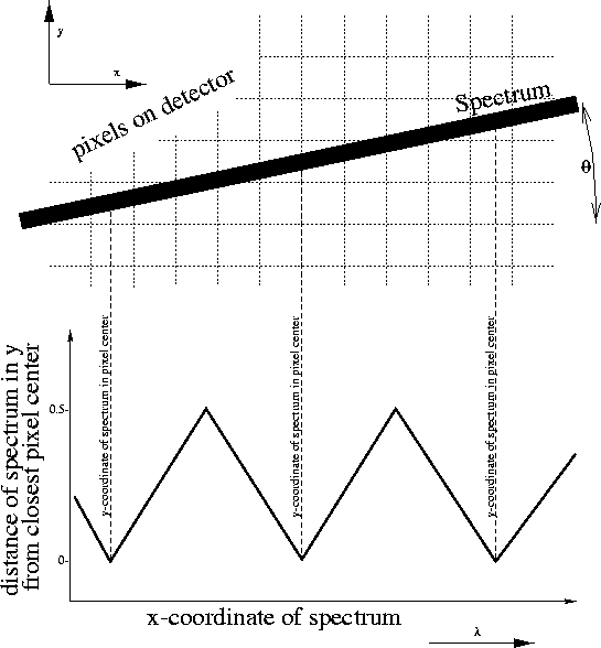
y-locations (see figure 3.11). If distortions in the
spectra are neglected, the y-position of a spectrum as a function of its
x position for a given inclination  can be written as
  , where
 and
and  are the coordinates of the spectrum on
the direct image, and
  is the offset between the grism
and direct image. The x position of the spectrum corresponds to the
wavelength of the extracted spectrum. Therefore, a non-zero
  implies that at some wavelength, the spectrum is located between two
rows on the detector, while at other wavelengths, the spectrum is
centered on a particular row.
Figure 3.11:
Schematic of the spectrum location as a function of
x-coordinate. The spectrum is inclined relative to the rows of the
detector by an angle  . This inclination puts the center of the
spectrum at different y-locations as a function of wavelength. The
spectrum is at the center of a row at the indicated positions, which
correspond to different wavelength wavelengths. A change of
  or and overall shift of the spectrum along the y-axis by a fraction of
a pixel will change the wavelength at which the centers of rows are
crossed.
 |
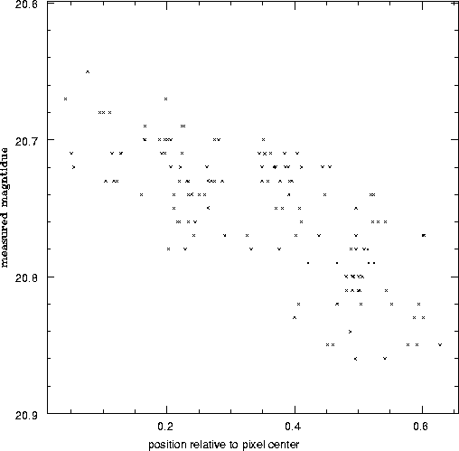
Figure 3.12:
Measured raw magnitudes of a star in the HDF-S field. The
magnitude of the same star has been measured on individual exposures,
which were taken at different dither positions. The magnitudes are
plotted as a function of the measured position of the star relative to
the closest pixel center. The data were kindly provided by Richard
Hook.
 |
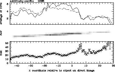
Figure 3.13:
A NICMOS Grism spectrum of the
flux calibrator P330e. The middle panel shows a cutout from the NICMOS
grism images. Wavelength decreases to the right. Note that at any
wavelength, most of the flux is contained in one or two pixels. The
regions where the spectrum lies between two rows can easily be
identified. At these places, the flux in the spectrum seem to be below
the flux in adjacent regions in the spectrum. This visual impression
is strengthened by the plot in the upper panel, which shows the
average of each column in the cut out as a function of x coordinates
relative to the position of the object on the grism image. Finally,
the lower panel shows a spectrum extracted by NICMOSlook with the PRF
correction option turned off. The x-axes of all three panels are
aligned. Comparison of the upper and lower panels show that the flux
calibration of NICMOSlook correctly converts the raw fluxes into the
expected overall shape of the spectrum. However, the flux is
underestimated where the spectrum crosses the boundary between rows.
 |
The quantum efficiency (QE) of NICMOS depends on the exact position
within each pixel where the photon hits the detector. The effect of
this intra-pixel response function (PRF)on the photometry of point
sources is shown in figure 3.12. A similar impact is
expected on the flux calibration of extracted spectra of point
sources. The flux in each wavelength bin of a spectrum is similar the
aperture magnitude measured in point source photometry. The
inclination of the spectrum relative to the rows implies that the
quantum efficiency and therefore the flux calibration of spectra
is a function of the intra pixel position of the object. This
is shown in Figure 3.13, which shows a grism image
and extracted spectra of a calibrator source. The spectrum is
modulated by a wave-like pattern. The phase of this pattern is
determined by the fact that the QE is at its minimum where the
spectrum falls between two rows.
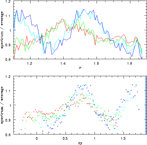
Figure 3.14:
Four spectra of calibrator P330e taken with grism G141. The four
spectra were placed at different dither positions, which resulted in
different sub-pixel locations of the object. The upper panel shows the
spectra as extracted by NICMOSlook, divided by a model of the spectrum
of the star. It can be seen that the different extracted spectra
deviate in a wave-like pattern with an amplitude of about 10% from
the average spectrum. The lower panel shows the same data plotted as a
function of  , where y is the row of
the spectrum location, and yi is an arbitrary integer number chosen
separately for each spectrum. It can be seen that the imprinted
pattern of the individual spectra are now shifted and are in phase. This
demonstrates that the phase of the pattern is determined by the
intra-pixel position of the spectrum.
 |
Figure 3.15:
Same plot as figure 3.14, but for grism G096.
The inclination  of G096 is larger than the one of G141. This
is the reason for the larger range of
  covered. In the
spectrum, this will show as a shorter period of the PRF pattern in
the spectrum.
 |
The effect on the flux calibration is shown in figures 3.14
and 3.15. The figures show extracted spectra of a
calibration source extracted from dithered exposures which lead to
different sub-pixel y-coordinates of the star. It can be seen that
the amplitude of the wave-like pattern imprinted on the spectra is
about 10%, the period is determined by the inclination of the
spectrum relative to the rows of the detector, and the phase is
determined by the intra-pixel position of the spectrum.


Next: Correction of Extracted Spectra
Up: Correction of Spectra for
Previous: Correction of Spectra for
Wolfram Freudling
5/29/1999
![]() can be written as
can be written as ![]() , where
, where
![]() and
and ![]() are the coordinates of the spectrum on
the direct image, and
are the coordinates of the spectrum on
the direct image, and ![]() is the offset between the grism
and direct image. The x position of the spectrum corresponds to the
wavelength of the extracted spectrum. Therefore, a non-zero
is the offset between the grism
and direct image. The x position of the spectrum corresponds to the
wavelength of the extracted spectrum. Therefore, a non-zero ![]() implies that at some wavelength, the spectrum is located between two
rows on the detector, while at other wavelengths, the spectrum is
centered on a particular row.
implies that at some wavelength, the spectrum is located between two
rows on the detector, while at other wavelengths, the spectrum is
centered on a particular row.
